원본 기사
기사
눈 스캔으로 신경퇴행성 질환 조기 진단 기술 개발
카타르의 라야즈 말리크 교수가 파킨슨병, 다발성 경화증, 치매 등 신경퇴행성 질환을 증상 발현 몇 년 전부터 감지할 수 있는 '각막 공초점 현미경' 기술을 개발했습니다.
각막 공초점 현미경 기술의 혁신
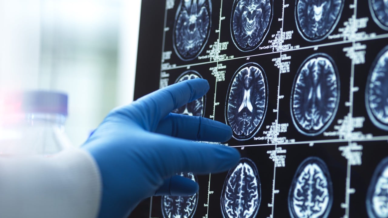
웨일 코넬 의과대학 카타르 및 하마드 의료법인의 라야즈 말리크 교수는 지난 25년간 각막 공초점 현미경(Corneal Confocal Microscopy, CCM)이라는 획기적인 눈 영상 기술을 개발해왔습니다. 이 기술은 파킨슨병, 다발성 경화증, 치매, 당뇨병성 신경병증 등 다양한 신경퇴행성 질환을 증상이 나타나기 수년 전에 감지할 수 있는 능력을 가지고 있습니다. 말리크 교수는 이 기술이 간단한 눈 스캔을 통해 2~3분 안에 환자의 눈에 있는 신경섬유의 이미지를 얻을 수 있다고 설명했습니다.
AI 기반 진단 및 미래 전망
수집된 신경섬유 이미지는 AI 알고리즘을 통해 분석됩니다. 이 AI는 신경섬유의 특징을 인식하고 손상 여부를 확인하여 질병 발생 위험을 예측합니다. 건강한 사람과 비교하여 손상된 신경섬유를 식별하는 방식으로 작동하며, 말리크 교수는 이를 통해 당뇨병성 신경병증은 증상 발현 5년 전, 치매는 3년 전부터 진단이 가능하다고 밝혔습니다. 말리크 교수는 현재 신경퇴행성 질환은 진단 시 이미 너무 늦은 경우가 많지만, 이 기술이 조기 경고 시스템으로서 질병에 대한 대처 방식을 바꿀 수 있을 것이라고 강조하며, 질병 진단 분야에 '눈의 빛'을 비추고 싶다는 포부를 전했습니다.
*출처: YouTube: Al Jazeera English (2026-05-12)*
관련 기사


📧 뉴스레터 구독
매일 아침 글로벌 뉴스 브리핑을 이메일로 받아보세요.
아직 무료입니다.